
What Is Keratoconus?
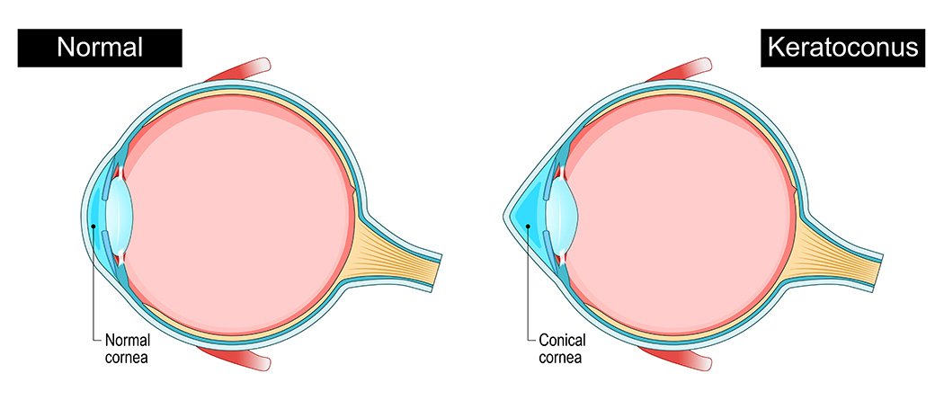
The cornea is the clear, dome-shaped surface at the front of your eye. In a healthy eye, it holds a fairly round shape. With keratoconus, that dome gradually thins and begins to bulge outward into more of a cone shape. That shift distorts the way light enters your eye, which is what causes the blurry, distorted, or double vision that many patients describe.
Keratoconus usually shows up in the teens or early twenties and progresses slowly over time, though the rate varies a lot from person to person. Some people experience only mild changes for decades. Others notice more rapid shifts, especially early on.
Common Signs Of Keratoconus
- Vision that blurs or distorts, even with a fresh glasses prescription
- Increased sensitivity to light or glare, especially at night
- Halos or streaking around lights
- Frequent prescription changes that don’t seem to stabilize
- Difficulty with contact lenses you previously wore without issue
How Keratoconus Is Diagnosed
A standard eye chart won’t catch keratoconus on its own. Diagnosis typically involves corneal mapping, which creates a detailed topographic image of your cornea’s surface. At Premier Eye Center, we use advanced diagnostic technology to detect corneal irregularities early, often before symptoms become disruptive.
If you’ve been bouncing between optometrists getting updated prescriptions without lasting improvement, that’s worth bringing up at your next visit. Keratoconus is one of those conditions that’s easy to miss if no one’s specifically looking for it.
Managing Keratoconus: What Are the Options?
Early on, updated glasses or soft contact lenses may be enough to manage mild keratoconus. As the condition progresses, specialty contact lenses become the more effective route, since they create a smooth optical surface over the irregular cornea.
Scleral Lenses
Scleral lenses are one of the most effective options for keratoconus patients, and they’re available right here in Prescott. These larger lenses vault completely over the cornea and rest on the white of the eye (the sclera). Because they don’t touch the cone at all, they’re often more comfortable than traditional rigid lenses, and they can provide significantly sharper vision.
Premier Eye Center was one of the first practices in the area to offer scleral lens fittings locally. Before that, patients had to drive to Phoenix for a series of appointments. We’re able to fit most patients in just a few visits, which makes the whole process a lot more manageable.
Learn More About Our Specialty Contact Lens Services
Why the Right Fit Matters
Fitting specialty lenses for keratoconus isn’t the same as a standard contact lens exam. It requires careful mapping of the cornea, a thorough understanding of how different lens designs interact with irregular surfaces, and often several visits to refine the fit. An ill-fitting lens on an already sensitive cornea can cause discomfort, corneal abrasion, or worsen the situation.
Our doctors have extensive experience fitting challenging corneas, including patients who’ve been told elsewhere that contacts simply won’t work for them. That’s a conversation worth having if you’re currently managing keratoconus on glasses alone.
Schedule an Appointment in Prescott
If you’re dealing with vision that doesn’t quite make sense, or if you’ve already been diagnosed with keratoconus and want to explore your options, we’d be glad to help. Premier Eye Center serves patients throughout the Prescott area, including Prescott Valley, Chino Valley, and Dewey-Humboldt.
Call us at (928) 778-3937 and schedule an appointment.
Living with Keratoconus
Keratoconus doesn’t have to mean accepting poor vision. Many people with the condition go on to drive, work, and live normally with the right lens solution and regular monitoring. The key is catching it, keeping an eye on it, and not settling for “good enough” when clearer vision is genuinely achievable.
If you have a family member with keratoconus, it’s worth knowing there is a genetic component, so siblings and children of someone diagnosed may benefit from screening as well.
Treating Keratoconus
A patient with early keratoconus was diagnosed during a routine vision exam and referred for a corneal cross-linking procedure to help prevent the progression of keratoconus.
Keratoconus (KC) is a progressive eye disease where the normally round, dome-shaped cornea gradually becomes thinner and irregular over time. As a result, people with keratoconus may notice blurry or distorted vision, increased light sensitivity, glare or halos at night, and frequent changes in their eyeglass prescription.
Keratoconus usually begins in the teenage years or early adulthood and can slowly worsen over time. In early stages, vision can often be corrected with glasses or soft contact lenses. As the condition progresses and the cornea becomes more irregular, specialized contact lenses like scleral lenses can achieve better vision than glasses.
Early detection of keratoconus is especially important today because of a treatment called corneal crosslinking (CXL). Corneal crosslinking is a minimally invasive procedure that strengthens the cornea helping it become more stable and less likely to continue bulging.
While crosslinking does not typically reverse keratoconus, it can stop or significantly slow the progression of the disease. Detecting keratoconus early allows doctors to treat patients before significant corneal damage occurs, preserving vision and reducing the likelihood that more invasive treatments—such as a corneal transplant—will be needed later in life. Because keratoconus can progress silently in its early stages, regular eye exams and modern corneal imaging technologies are key to identifying the condition early.
