Advanced Diagnostic Procedures2026-08-19T08:08:20-07:00

Advanced Diagnostic Testing at Premier Eye Center

Not every eye problem is obvious.
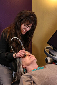
dry-eye-wesWhen a routine exam turns up something that needs a closer look, Premier Eye Center has the diagnostic tools to follow through — right in the same office. Advanced imaging can detect early signs of glaucoma, macular degeneration, and diabetic changes before they affect your vision. Electrodiagnostic testing evaluates the visual pathway for neurological conditions.

Corneal mapping supports contact lens fitting and keratoconus monitoring. Peripheral vision screening is included with every comprehensive exam at no extra charge.

Whatever the equipment, your doctor explains the results before you leave.

Click Here & Schedule An Appointment Today

Common Questions About Comprehensive Eye Exams

What Makes a Test “Advanced Diagnostic”?2026-06-26T11:50:15-07:00

A comprehensive eye exam covers a lot of ground — refraction, binocular vision testing, tonometry, a retinal exam, and a screening visual field. That’s where we start for every patient. When findings from that exam raise a question — a suspicious optic nerve, unusual corneal curvature, abnormal pressure readings, early retinal changes — that’s when we turn to the instruments below.

These tools produce data that can’t be gathered with a slit lamp and a focusing lens alone. Some of them measure tissue in micrometers. Some capture electrical responses in your retina and visual cortex. Most are completely painless and take just a few minutes.

Do I need a referral to have these tests done?2026-06-26T11:47:30-07:00

No. You can schedule directly with Premier Eye Center. If your exam or a specific symptom warrants advanced testing, your doctor will order it during your visit.

Will I get my results the same day?2026-06-26T11:47:14-07:00

Yes. Our doctors review and explain diagnostic results during your appointment. You won’t leave wondering what the images showed or what the numbers mean.

How long does advanced diagnostic testing take?2026-06-26T11:46:57-07:00

Each test is relatively brief — most take two to five minutes. If your exam includes multiple diagnostic procedures, the overall appointment may run longer than a standard exam. Our front desk can give you a realistic time estimate when you schedule.

Do any of these tests require dilation?2026-06-26T11:46:40-07:00

Most of the tests described on this page do not require dilation. OCT imaging, visual field testing, corneal topography, electrodiagnostic testing, and pachymetry are all performed without dilating drops. Your doctor will let you know in advance if dilation is needed for any part of your exam.

Do these tests cost extra?2026-06-26T11:46:22-07:00

Most advanced diagnostic tests are ordered when clinically indicated and are billed to medical insurance, not vision plans. The Humphrey Visual Field screening is included in every comprehensive exam at no additional charge. If advanced testing is recommended during your visit, your doctor will discuss what’s involved and how it’s billed. We accept most major medical insurance plans as well as vision plans — see our insurance page for details.

The Diagnostic Instruments We Use

Age-related macular degeneration (AMD) is one of the leading causes of permanent vision loss in adults over 60. What makes it particularly difficult to manage is that by the time most patients notice symptoms — blurry central vision, distortion, difficulty reading — the disease has often been progressing for years.

The AdaptDx Pro tests dark adaptation, which is the eye’s ability to recover its sensitivity after exposure to bright light. This function degrades in the early stages of AMD, often years before structural changes show up on imaging. Our doctors use it to detect macular degeneration in its earliest stages, when there’s the most opportunity to slow its progression through management and monitoring. If you have a family history of AMD, are over 60, or have other risk factors, this test can be an important part of your exam.

The Diopsys Nova is one of the more sophisticated pieces of equipment in our practice. It performs two distinct types of electrodiagnostic testing: Visual Evoked Potential (VEP) and Electroretinogram (ERG).

ERG (Electroretinogram) measures the electrical response of the retina — specifically, how the photoreceptor cells respond to light stimulation. This gives our doctors an objective picture of retinal function that doesn’t rely on the patient describing what they see. ERG testing has helped us diagnose glaucoma earlier than conventional methods allow, rule it out in ambiguous cases, and in some patients, has supported discontinuing medication when test results showed the retina was functioning normally.

VEP (Visual Evoked Potential) measures how the visual signal travels from the retina through the optic nerve to the visual cortex in the brain. It’s useful for evaluating optic neuritis and managing patients with neurological conditions such as multiple sclerosis, where the optic nerve is often affected.

Both tests are objective — no verbal response or button-pushing required. You look at a pattern on a screen a few feet away while the instrument records the electrical signals. It’s quiet, fast, and completely non-invasive.

Testing that was once only available at university research centers and specialty hospitals is now available here in our Prescott office.

OCT stands for Optical Coherence Tomography. Our Optovue iVue is a Spectral Domain OCT that captures detailed cross-sectional images of the retina and optic nerve at 26,000 A-scans per second. To put that in practical terms: it produces highly detailed structural maps of tissue layers you can’t see with any other clinical instrument.

We use the iVue to detect the earliest structural signs of glaucoma (thinning of the retinal nerve fiber layer), macular degeneration (drusen, fluid, and tissue changes at the macula), and diabetic macular edema (swelling in the central retina). The scans are stored and compared to your previous visits, so subtle year-over-year changes don’t go unnoticed.

Testing is painless and takes less than two minutes. No dilation is required. Your doctor reviews the results with you the same day.

The Sonogage pachymeter uses ultrasound to measure corneal thickness in micrometers — it’s one of the most precise measurements we take in the office.

We use it primarily in two situations. For glaucoma management, corneal thickness is a meaningful factor in interpreting eye pressure readings. Research has confirmed that thin corneas are associated with increased risk of glaucoma progression, and a pressure reading from a thin cornea can underestimate the actual intraocular pressure. Knowing corneal thickness helps our doctors interpret tonometry results more accurately.

For patients exploring LASIK or other refractive procedures, pre-operative corneal thickness is a critical data point. LASIK uses a laser to reshape corneal tissue, which means the cornea needs enough baseline thickness to remain structurally stable after surgery. We take this measurement as part of LASIK co-management to help patients and their surgeons make well-informed decisions.

High-resolution retinal photographs give us a permanent, dateable record of your retina’s appearance. That record becomes valuable over time, when we can compare current images to past ones to spot gradual changes.

Our Canon imaging system uses a low-intensity flash to minimize discomfort. The Synemed software lets our doctors magnify specific areas, invert color contrast to highlight subtle details, and track changes across multiple visits. Being able to show a patient their own retinal images — and explain exactly what’s changed and what hasn’t — also helps make an often-abstract diagnosis feel concrete and understandable.

The Humphrey Visual Field Analyzer is the accepted standard for measuring peripheral vision in clinical and research settings. Visual field loss is a hallmark of glaucoma, but it’s also associated with strokes, macular degeneration, neurological conditions, and other disorders affecting the visual pathway.

Every comprehensive eye exam at Premier Eye Center includes a peripheral vision screening at no additional charge. The screening takes approximately 90 seconds per eye. If that screening suggests an area worth investigating further, your doctor may order a threshold visual field test, which creates a more detailed map and provides the kind of progressive data needed to monitor change over time.

Northern Arizona’s climate — dry air, high altitude, low humidity year-round — makes dry eye syndrome more common here than in much of the country. After a full diagnostic workup for dry eye, our doctors may recommend intracanalicular punctal plugs as part of a treatment plan.

For patients who haven’t tried plugs before, we offer a diagnostic option: small temporary plugs placed in the tear drainage canals that allow you to experience a four-to-seven-day trial period without any long-term commitment. The plugs dissolve on their own after the trial period. If your symptoms improve during that window, our doctors will discuss longer-lasting plug options or other interventions — including our dry eye treatment program using InMode Forma-I and Lumecca-I technology — to provide ongoing relief.

The reason we have these instruments isn’t just to generate data. It’s to catch problems early enough to make a difference, and to manage conditions with the precision they require.

The Premier Eye Center are licensed and trained to treat ocular disease — not just detect it. If advanced testing reveals elevated glaucoma risk, early macular changes, or corneal abnormalities, you won’t be handed a referral and sent to start over somewhere else. We manage these conditions in our practice, often from first detection through long-term monitoring and treatment. When a surgical subspecialist is genuinely needed, we maintain close working relationships with ophthalmologists and other specialists in the region.

For a full list of conditions we diagnose, treat, and co-manage, see our Treatment of Ocular Disease page.

Go to Top